A ‘muscle man’ from De humani corporis fabrica, annotated by a reader.Credit: Bibliotecha Civica Romolo Spezioli, Fermo. Call no.: 1e7n.1582
The Renaissance anatomist Andreas Vesalius’s De humani corporis fabrica (‘On the fabric of the human body’) is a foundational work of medicine in the West. Its more than 200 woodcuts revolutionized how people pictured the human body, flayed and cut to reveal musculature, nerves, organs and bones. Even now, 475 years after it was first published, the bold images of skeletons and skinless ‘muscle men’ in sinuous poses (by illustrator Jan Steven van Calcar) beguile.
More than 700 copies survive from the 1543 and 1555 editions, which Vesalius supervised. Of these, roughly two-thirds contain comments in the margins, bizarre doodles, and coloured-in and even defaced images, as we reveal in our book The Fabrica of Andreas Vesalius. Early readers, on evidence, studied Vesalius’s treatise diligently, yet had no compunction about scribbling in a hugely expensive volume.
Looking deeper, the marginalia tell two stories. One is that some found the images baffling, and attempted to clarify them in innovative ways. Another is that the pious found the figures’ necessary nudity scandalous, and felt impelled to weigh in with ink and scissors. Our study of the reactions of hundreds of readers has taught us that medical communities do not always adopt innovative solutions quickly, even when they are presented in such an elegant format as the Fabrica. It takes time to get used to novelty. And we have learnt that even the most ingenious scientific minds can fail to predict how political and religious institutions will respond to their work.

The portal vein, with the haemorrhoidal vein coloured in.Credit: The Provost and Fellows of The Queen’s College, Oxford
The Fabrica’s early readers were the first generation of physicians and surgeons in Europe to face the daunting task of using detailed printed images to identify the organs of the body and learn about human physiology. Vesalius and van Calcar faced challenges of their own. The Fabrica’s image of the branching portal vein, which carries blood from the intestines to the liver, is highly complex — and does not quite succeed. It is almost impossible, for example, to single out the haemorrhoidal vein. (At the time, this was important because the vein was supposed to be the cause of both menstruation and haemorrhoids, thought to be analogous processes that purged corrupted blood from the body.) Thus, in a copy now in the library of Queen’s College at the University of Oxford, UK, someone used a quill and red ink to colour in this meandering vein, like a child playing a maze game.
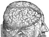
In a copy once owned by Nuremberg physician Georg Palma, an intricate image of the brain is ‘enhanced’. Palma painted six pairs of cranial nerves different hues in watercolour and used the same colours to underline the corresponding pairs in the text on the following page.

An image of the brain with cranial nerves colour-coded in pairs.Credit: Stadtbibliothek im Bildungscampus Nürnberg, Med. 155.2°, p. 511 and 513
Even Vesalius realized that his images could be confusing, and devised an ingenious method to explain them. A letter or number was printed onto the image of each body part, with a separate key. Unfortunately, the characters were often too small to pick out against the swirling background. Some frustrated readers underlined, highlighted, enlarged or repeated the characters in the margins. On one muscle man, for instance, the tiny character identifying a thigh muscle was barely visible, and a confused reader queried desperately whether it was the Greek letter µ or the Roman letter u.
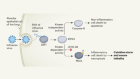
Faced by such challenges, many medics might have given up on the images. Indeed, when we reconstructed what early modern readers and scholars found fascinating about the Fabrica, it was evidently the text. The clear majority of sixteenth- and seventeenth-century readers who annotated the book focused on that and left no traces of having engaged with the illustrations. Sixteenth-century reviews of the Fabrica confirm this impression, because they tended to discuss only the text.
This is no surprise. The Fabrica’s scholarly readership was trained in the traditions of Renaissance humanism, which put a strong emphasis on textual analysis. Even if they found it difficult to interpret visual information, medical practitioners were expert at making sense of long Latin texts. Furthermore, the body’s ‘interior universe’ had hardly been mapped. Even today, it is difficult to make sense of images of internal organs if you’ve never seen a dissected body, and radiologists need years of training to interpret X-rays or magnetic resonance imaging scans.

An illustration of the male torso censored with a ‘modesty apron’.Credit: Bibliotheque municipale de Bourges, Bibliotheque des Quatre Piliers. Call no.: B 2338
If images were not that helpful for understanding the body, what was their purpose? For Church authorities in the period, the answer was clear. They argued that such figures held an erotic appeal because they showed the genitals — and so should be censored. The first version of the Index librorum prohibitorum, the list of books banned by the Catholic Church, came out in 1559, and it did not mince words about ‘licentious’ books. That included tomes on anatomy. Many owners of the Fabrica felt that they had to paint aprons on the muscle men (as in the copy once owned by the Jesuit College of Bourges in France), or snip the offending parts out.
Only a minority of copies of the Fabrica were so treated. We checked every surviving copy, and found the images intact in the majority owned by Catholics in the period. That invasive censorship happened at all signals that, at least until the trials of Galileo Galilei in the early seventeenth century, the Church found anatomical illustrations more dangerous than heliocentrism.
 The man who bared the brain
 Leonardo's anatomy years